Summary
What are chromosome studies?
Chromosome studies, also known as karyotyping, is a test in which a specialist pathologist or scientist looks at the number and structure of chromosomes. They look for changes that are known to cause any of a range of different health conditions.
A quick look at genes and chromosomes
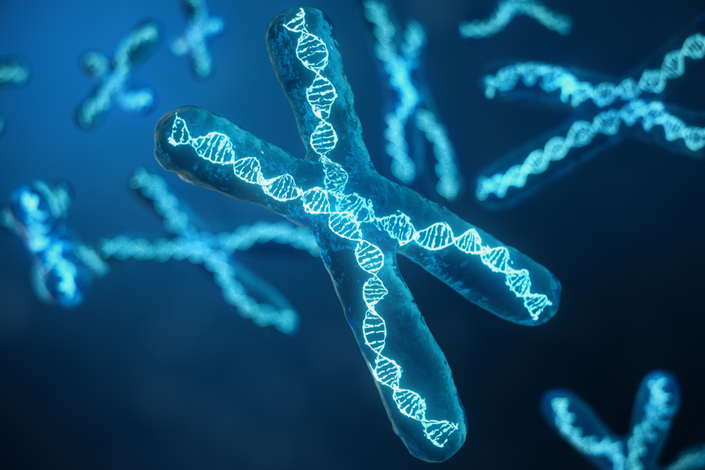
Our bodies are made up of trillions of cells. Almost every cell has a nucleus (a sac in the middle of the cell) containing a complete set of genetic material in genes. Genes are the chemical instructions that tell your body how to make all the different parts it needs to function. Genes are short sections of DNA and our DNA is packaged into structures called chromosomes
Usually, our cells have a set of 23 pairs of chromosomes or 46 individual chromosomes. There are 22 pairs of chromosomes which are the same in both males and females, these are called autosomes, and one pair of sex chromosomes - a pair of X chromosomes in females (XX) and one X and one Y in males (XY).

How genes and chromosomes are passed from parents to children
Chromosomes are inherited from our parents. We get one copy of each chromosome from our mother (22 autosomal chromosomes and an X chromosome, via the egg), and another copy of each chromosome from our father (22 autosomal chromosomes and either an X or a Y chromosome, via the sperm).
While typically cells have 46 chromosomes, sperm and egg cells are different. They have only 23 chromosomes. When a sperm cell and an egg cell fuse in conception, they each bring 23 chromosomes to form a fertilised egg that has a full set of 46 chromosomes. Combining genetic information from both parents in this way ensures genetic diversity.
Chromosomal changes
Chromosomal changes can occur when the egg or sperm cells are made inside a person’s body, or in the early stages of conception after an egg has been fertilised. When this happens, these changes (called genetic variants or mutations) are present in every or nearly every cell in a person’s body. These are called germline genetic variants.
Chromosomal changes can also occur during the normal course of life. Our cells have a lifespan – they get old and die off. Some cells last only a few hours or days, while others last for months or years or even a lifetime. The cells in our bodies are constantly dividing to make new cells. Each time a cell divides, it copies its DNA and sometimes small copying mistakes can occur, called variants. Some cells (especially cancer prone ones) have unstable chromosomes that can break and rejoin in the wrong way when the cell is copying itself. These are called acquired or somatic genetic variant.
Summary
Types of chromosomal changes
When changes happen, whole chromosomes may be lost or added, meaning a person is born with one more or one less chromosome than the usually 46 chromosomes. Other changes can happen when pieces of a chromosome are rearranged within one chromosome or transferred between two or more chromosomes. Small pieces of a chromosome may be lost, or an extra piece added, resulting in too little or too much genetic information. Some of these changes can cause health and developmental problems, while others may have no effect on a person's health.
The way these chromosomal changes impact on our health is very varied and depends on which chromosome has been changed and in what way.
Why get tested
Chromosome studies or karyotyping is used to:
How are chromosome studies performed?
Chromosomes are extremely small and cannot be seen other than when cells are dividing.. When a cell prepares to divide, the DNA tightly coils and condenses, forming visible chromosomes. This process makes the chromosomes visible and helps prevent them from getting tangled or breaking as they are moved during cell division. At this time, they can be viewed under a microscope.
Chromosome pairs are numbered from 1 to 22, with the 23rd pair labelled as XX or XY.
Karyotyping is performed by:
Each chromosome looks like a striped straw. It has two arms that differ in length. These are a short arm (p) and a long arm (q). The pinched-in area between the arms is called a centromere.
Each chromosome will show up as a series of light and dark horizontal bands. The length of the arms and the location of the bands help show which is the top and which is the bottom.
Examples of different karyotypes


Having the test
Sample
Different samples may be needed depending on the reason for testing. Testing is usually done with a blood sample especially when testing for germline variants. However, a sample of your bone marrow or a tissue sample taken in a biopsy procedure may be needed for some conditions.
If a sample if being collected to test an unborn baby, a sample of the placenta will be taken via a procedure called Chorionic Villus Sampling (CVS) or a sample of the amniotic fluid (the fluid surrounding the baby is in when in the womb) will be taken via a procedure called an amniocentesis.
The genetic makeup of the cells of the placenta are usually the same as the genetic make up of the cells of the baby (although, in rare cases, this may not be the case) and so testing these cells with a CVS is usually the same as testing the genetic make-up of the baby. The amniotic fluid contains skin cells that flake off the baby as it grows in the womb, and these are tested when the amniotic fluid is collected.
Any preparation?
None.
Your results
Each chromosome contains hundreds or thousands of genes in specific locations. The bigger chromosomes have more genes than smaller chromosomes.
Changes can affect the number and the make-up of chromosomes. Anything other than a complete set of 46 chromosomes represents a change in the amount of genetic material present and can cause health and development problems. A change in how the genetic material on the chromosomes is arranged, without changes to the amount of genetic material present, can also cause health issues.
Where there are structural changes, (when the position of the genetic material on the chromosome changes but the amount of genetic material remains the same), the severity of problems varies depending on the chromosome. Also, the effect on health may vary from person to person, even when the same chromosome changes are present.
Reading your test report
Chromosome reports can be very complex and need to be carefully interpreted. You should discuss your results with a doctor or genetic counsellor. Only the simplest examples are described here.
Results you may see on your chromosome report
| Result | What it means |
| 46,XX | This is a karyotype with XX sex chromosomes. It represents the usual chromosome make-up for a biological female.
|
| 47,XX,+21 | This is a karyotype with XX sex chromosomes which has a whole extra copy of chromosome 21, as seen in Down syndrome.
|
| 46,XY,dup(1)(q22q25) | This is a karyotype with XY sex chromosomes which has a total of 46 chromosomes. There is an extra piece (dup, meaning duplication) on chromosome 1 which sits between band 22 and band 25 on the long arm (q). (Bands are specific locations on chromosomes.) Deletions (missing bits – “del”) or insertions and duplications (extra bits) are described referring to the chromosome affected, and the position of where the change is seen. |
Terms you may see on your report
| Term | Meaning |
| Trisomy | There is a whole extra chromosome, so there are three copies instead of two. Conditions associated with trisomies include Down syndrome (trisomy 21), Patau syndrome (trisomy 13) and Edward syndrome (trisomy 18). |
| Monosomy | A chromosome is missing. An example of monosomy is Turner syndrome (a female with a single X chromosome – X instead of XX). Most other monosomies are not compatible with life. |
| Deletions | There are missing pieces of chromosomes and/or genetic material. Some may be small and can be difficult to be detected. |
| Duplications | There is extra genetic material, and this may be on any chromosome, for example, two horizontal bands at a specific location instead of one. |
| Translocations | Pieces of a chromosome break off and reattach to another chromosome (called a rearrangement). Translocations can be balanced or unbalanced. A balanced translocation is a rearrangement where segments from two different chromosomes swap places without any gain or loss of genetic material. This means the person will not have an extra or any missing genetic material, but the genetic material is in a different place to where it is usually. People with balanced translocations are not usually affected, but it may lead to issues with fertility, recurrent miscarriage or health issues in their children as their children may inherit an unbalanced form of their translocation. An unbalanced translocation is where genetic material is gained or lost between chromosomes. This results in the person having too much or too little genetic material, which can affect development and health. |
| Inversions | A segment of a chromosome breaks in two places, flips around and reattaches to the same chromosome in the opposite direction. DNA may or may not be lost in the process. |
| Mosaicism | It is possible for people to have cells in their body with differing genetic material. This happens when genetic variations happen early in the development of a fetus, leading to that person having some cells with one type of genetic makeup and some cells with another type of genetic makeup. This is called mosaicism or mosaic genetic variation. For example, in some cases of Down syndrome a person can have some cells with an extra third copy of chromosome 21, and some cells with two copies of chromosome 21. Generally speaking, a person with mosaic genetic changes will not necessarily have less severe issues or symptoms than a person who has the genetic change in every cell in their body. However, in some disorders, mosaicism may mean symptoms are milder. |
Questions to ask your doctor
The choice of tests your doctor makes will be based on your medical history and symptoms. It is important that you tell them everything you think might help.
You play a central role in making sure your test results are accurate. Do everything you can to make sure the information you provide is correct and follow instructions closely.
Talk to your doctor about any medications you are taking. Find out if you need to fast or stop any particular foods or supplements. These may affect your results. Ask:
More information
What is Pathology Tests Explained?
Pathology Tests Explained (PTEx) is a not-for profit group managed by a consortium of Australasian medical and scientific organisations.
With up-to-date, evidence-based information about pathology tests it is a leading trusted source for consumers.
Information is prepared and reviewed by practising pathologists and scientists and is entirely free of any commercial influence.