What is being tested?
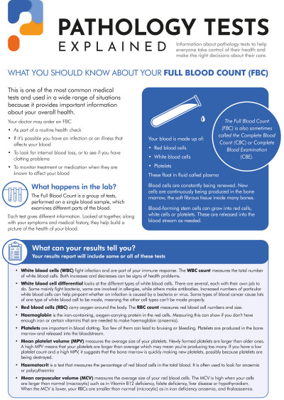
A blood film examination allows the evaluation of white blood cells (WBCs, leucocytes), red blood cells (RBCs, erythrocytes), and platelets (thrombocytes). These cell populations are produced and mature in the bone marrow and are eventually released into the bloodstream as needed. WBC’s main function is to fight infection, while RBCs carry oxygen to the tissues. Platelets appear as small cell fragments and, when activated, form a plug as one of the first steps in blood clotting. The number and type of each cell present in the blood is dynamic but generally maintained by the body within specific ranges. Values can fluctuate at times of illness or stress; intense exercise or smoking can also affect cell counts.
A peripheral blood film examination is a snapshot of the cells that are present in the blood at the time that the sample is obtained. To create a blood film, a single drop of blood is spread in a thin layer across a glass slide, dried, and then stained with a special dye. Once the stain has dried the slide is evaluated under a microscope by a medical scientist or haematologist.
The drop of blood on the slide contains millions of RBCs, thousands of WBCs, and hundreds of thousands of platelets. Under the microscope, the stained WBCs can be easily seen and counted to estimate the number of each type of cell present. In addition, one can compare their size, shape and general appearance to the established appearance of “normal” cells. It is possible to distinguish between the five different types of WBCs and to determine their relative percentages by counting 100 consecutive cells. During this examination, one can also evaluate the size, shape and colour (indicators of haemoglobin content) of the RBCs and also estimate the number of platelets present.
How is it used?
A peripheral blood film was once prepared on nearly everyone who had a full blood count (FBC) performed. With the automated blood cell counting instruments currently used, an automated WBC differential is also provided. However, if the presence of abnormal WBCs, RBCs or platelets is suspected, a blood film examined by a trained professional is still the best method for definitively evaluating and identifying immature and abnormal cells.
There are many diseases, disorders and deficiencies that can have an effect on the number and type of blood cells produced, their function and their lifespan. Although usually only normal mature cells are released into the bloodstream, circumstances can force the bone marrow to release immature and/or malformed cells into the circulation. When a significant number of abnormal cells are present, they can suggest an underlying condition and prompt the doctor to do further testing.
When is it requested?
A blood film examination is primarily ordered to evaluate blood cell populations when a FBC with WBC differential, performed with an automated blood cell counter, indicates the presence of abnormal or immature cells. It may also be performed when a doctor suspects a deficiency, disease or disorder that is affecting blood cell production, such as an anaemia, decreased or abnormal production of cells in the bone marrow, or increased cell destruction. A blood film examination may also be ordered when a patient is being treated or monitored for a blood cell-related disease.
What does the result mean?
Findings from the blood film evaluation are not always diagnostic in themselves and more often indicate the presence of an underlying condition and its severity and suggest the need for further diagnostic testing. Blood film findings may include:
RBC (Red blood cells)
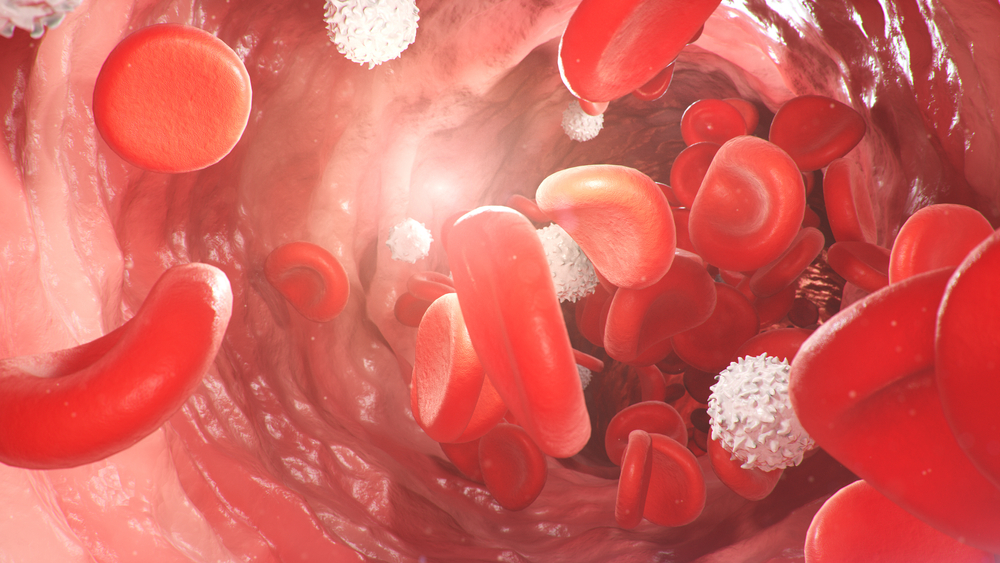
Normal, mature red blood cells are uniform in size (7 µm) and do not have a nucleus as most other cells do. They are round and flattened like a doughnut with a depression in the middle instead of a hole (biconcave). Due to the haemoglobin inside the RBCs, they appear pink to red in colour with a pale centre with routine staining. While not every RBC will be perfect, any significant number of cells that are different in shape or size may indicate a more severe problem. There may be one or more irregularities present and may include:
WBC (White Blood Cells)
White blood cells have a nucleus surrounded by cytoplasm. All WBCs are derived from bone marrow stem cells. In the marrow, they differentiate into two groups: myelocytic and lymphoid cells. They then mature into five distinct types of WBCs.
Platelets
These are cell fragments that derive from large bone marrow cells called megakaryocytes. Upon release from the bone marrow, they appear as fragments in the peripheral blood. When there is blood vessel injury or other bleeding, the platelets become activated and begin to clump together to form aggregates which is the beginning of a blood clot. You must have a sufficient number of platelets to control bleeding. If there are too few, the ability to form a clot becomes impaired and can be a life-threatening situation. In some people, too many platelets may be produced, which may result in interferences with the flow of blood, increasing a person's risk of developing a blood clot. These same people may also experience bleeding because many of the extra platelets may be dysfunctional even though they appear normal.
Enumeration of platelets is usually part of a FBC. An abnormally low number or high number of platelets may be further evaluated by preparing a peripheral blood film to directly visualise any anomalies in shape, size or granularity.
Is there anything else I should know?
Some examples of situations or conditions that may affect or invalidate results of a blood film examination include:
Common questions
It has on a routine basis, but the automated blood cell counter usually evaluates the RBCs, WBCs and platelets based on their shape, size, and electrical or photometric properties. There can be some variation in each cell type and numbers the body produces due to a variety of physiological and external stimuli. Use of an automated instrument can often identify the presence of abnormal cells but lacks the ability to definitively subclassify them. Cell fragments and platelet clumps, particularly if they are large in size, can be mistakenly counted as WBCs, thus falsely elevating a white cell count. A medical scientist or haematologist can see these abnormalities on a blood film and has been trained to identify and classify them appropriately.
More information
Pathology Tests Explained (PTEx) is a not-for profit group managed by a consortium of Australasian medical and scientific organisations.
With up-to-date, evidence-based information about pathology tests it is a leading trusted sources for consumers.
Information is prepared and reviewed by practising pathologists and scientists and is entirely free of any commercial influence.